Introduction
Inge Endler, widely considered the father of Echocardiography, revolutionised the field in 1953 with the introduction of this tool. Since then, it has allowed radiologists and cardiologists to get an in depth view of the beating heart, non invasively and to diagnose multiple, potentially life threatening cardiac conditions so that timely intervention may be performed and lives saved.
This tool is also very frequently used in the paediatric population, to screen and diagnose for heart diseases affecting children from birth.
Our Purpose
We are a team of a Paediatrician in training and a well renowned Pediatric and Fetal cardiologist in Mumbai.
We, as doctors also saw the need that training of physicians would benefit their holistic knowledge, and also allow for better daignosis of patients.
One of the simplest would be in the management of a dehydrated child. It is sometimes difficult to assess, when giving fluids to such a child whether one has overhydrated a child, which would put unneccessary load on the heart and worsen the situation. A simple sonography could be used to assess the distensibility of the great vein (Inferior vena cava), and if it is collapsing, that would be a green signal that we can still give the dehydrated and fluid depleted child more fluids. It also allows us to know the pumping efficiency of the heart.
Unfortunately, even though sonology machines are available to doctors at centres, very few have been trained on how to use them, much owing to the lack of patients who'd cooperate, and especially time constraints in training regimens.
Hence we decided it was time we built a simulator so as to teach doctors in training how to use a sonology machine to conduct a basic Echocardiography examination.
Our Plan
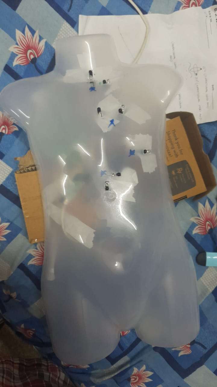
Our project involves a simple dummy, where a "probe" can be placed, and it would display the corresponding video image on screen one would expect in a child.
The doctors would then be instructed on proper probe placement, and how to interpret the displayed image.
The program also allows multiple views, so as to simulate different structural cardiacs defects such as a ventricular septal defect or a narrowed aortic arch, etc. These are basics a paediatrician would find to be relevant to their practice.
How the magic happens
We use a Raspberry-Pi B, which has a program coded in python. This Pi accepts input from multiple proximity sensors through it's GPIOs.
When the "probe" object triggers any of the proximity sensors, it plays the corresponding video.
Each proximity sensor is placed on the simulation dummy as would one expect the standard 2D Echocardiography probe placements in real life.
Our future plans are to increase the number of sensors, including addition of a tilt-gyroscope for more depth and realism of the simulations.